Quick answer: Finding a breast lump or fibroadenoma is frightening, but most breast lumps — especially in women under 35 — turn out to be benign, and fibroadenoma is the single most common cause. The only way to know for certain, though, is not by how a lump feels, but through a structured diagnostic process called triple assessment: clinical examination, imaging (ultrasound or mammography), and, when needed, a non-surgical tissue diagnosis using image-guided core needle biopsy — available at CVIC Indore (Center Of Vascular And Interventional Care). This guide explains exactly how that diagnostic pathway works, what imaging actually shows, and when a lump needs a closer look.
Few moments cause as much immediate anxiety as discovering a new lump in the breast. The mind understandably jumps to the worst-case scenario. The reassuring reality is that the large majority of breast lumps, particularly in younger women, are benign — and fibroadenoma, a non-cancerous overgrowth of glandular and connective tissue, is the most common benign breast lump diagnosed in women in their teens, twenties, and thirties.
But reassurance without evidence isn’t good medicine, and “it’s probably nothing” is not an adequate answer to give — or receive — about a breast lump. This article focuses specifically on the question most general breast-lump guides skip over: how does a doctor actually know whether a lump is a fibroadenoma or something that needs urgent attention? The answer lies in a structured, evidence-based diagnostic process — and increasingly, that process can be completed entirely without surgery, through image-guided core needle biopsy at CVIC Indore.
Breast Lump or Fibroadenoma: What Are We Actually Talking About?
A “breast lump” is simply any area of breast tissue that feels different, firmer, or more defined than the surrounding tissue. Not all lumps are the same kind of thing:
- Fibroadenoma — a benign, solid mass of glandular and fibrous tissue. Typically smooth, firm, rubbery, and mobile (“slippery” under the fingers), most common in women aged 15–35, often hormonally responsive (can enlarge during pregnancy or with hormonal contraception, and often shrinks after menopause).
- Cysts — fluid-filled sacs, often tender, that can fluctuate in size with the menstrual cycle.
- Fibrocystic changes — generalized lumpiness and tenderness related to hormonal fluctuation, not a single discrete lump.
- Lipomas — soft, benign fatty lumps.
- Malignant (cancerous) masses — typically, though not always, firmer, less mobile, and irregular in shape, and more likely to be painless in early stages.
The overlap in how these can feel on self-examination is exactly why touch alone is not a diagnostic tool.
Who Tends to Get Fibroadenomas?
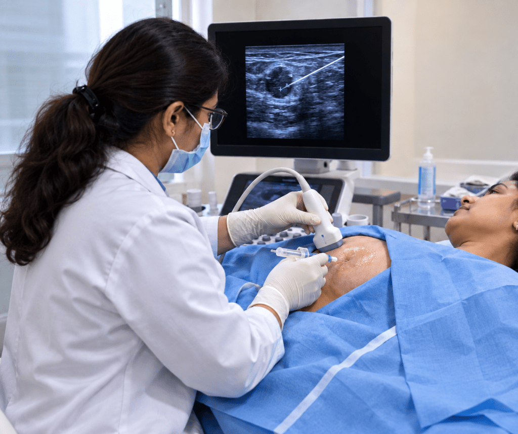
Fibroadenomas are the most common benign breast tumor in women under 35, and they are strongly linked to hormonal activity — which is why they are diagnosed most often during the reproductive years, can grow larger during pregnancy or while breastfeeding, and often shrink or calcify after menopause once estrogen levels decline. Regular self-awareness of how your own breast tissue normally feels remains genuinely useful — not to self-diagnose, but to notice a new lump or a change in an existing one early enough to have it properly evaluated, which is exactly where the diagnostic process below comes in.
Can You Tell Fibroadenoma From Cancer Just By How It Feels?
Honestly — no, not reliably, and this needs to be said plainly rather than glossed over. While classic teaching describes fibroadenomas as smooth, round, and mobile, and cancers as hard, irregular, and fixed to surrounding tissue, real-world presentations overlap more than most patients (and even some clinicians relying on touch alone) expect. Pain is a particularly unreliable indicator — most breast cancers are painless in their early stages, so the presence or absence of discomfort tells you very little on its own. Some malignant tumors, particularly in younger women, can feel deceptively smooth and mobile early on, closely mimicking a fibroadenoma.
This is precisely why a structured diagnostic process exists, rather than relying on clinical impression alone.
The Triple Assessment: How Breast Lumps Are Actually Diagnosed
The internationally recognized standard for evaluating a palpable breast lump is called triple assessment, and it is built on the principle that no single method — not examination, not imaging, not even biopsy alone — should be trusted in isolation. The three components are:
- Clinical examination — a doctor’s physical assessment of the lump’s size, shape, mobility, texture, and any associated skin or nipple changes, along with a review of personal and family history.
- Imaging — ultrasound and/or mammography, to characterize the lump’s internal architecture, something touch can never reveal.
- Tissue diagnosis — cytology (fine needle aspiration) or, more definitively, histology via core needle biopsy, examining actual cells or tissue under a microscope.
Each component is typically scored, and when all three point the same direction — especially when all three agree the finding is benign — diagnostic accuracy approaches 100%. It’s when these three components disagree, or when any one of them raises concern, that further investigation (often biopsy, even if imaging looked reassuring) becomes essential. This is the actual clinical logic behind “getting it checked” — it isn’t a single test, but an agreement across three independent forms of evidence.
Imaging: What Fibroadenoma Looks Like — and What Raises Concern
This is the part of the puzzle that a physical exam alone cannot provide, and it’s worth understanding in some detail, because it’s exactly what a radiologist is looking for when reading your ultrasound or mammogram.
On Ultrasound
| Feature | Typical Fibroadenoma | Features Raising Concern |
|---|---|---|
| Shape | Oval | Irregular |
| Orientation | Wider-than-tall | Taller-than-wide |
| Margins | Circumscribed (well-defined) | Spiculated or indistinct |
| Internal texture | Homogeneous (uniform), hypoechoic | Heterogeneous |
| Blood flow (Doppler) | Minimal | Increased, disorganized |
| Posterior shadowing | Absent or mild edge shadow | Present behind the mass |
On Mammography (typically added after age 40, or when clinically indicated)
Fibroadenomas classically appear as round, oval, or gently lobulated masses with smooth, circumscribed margins. Older fibroadenomas that have begun to calcify can develop a distinctive “popcorn-like” coarse calcification pattern — considered a reassuring, classic benign sign when present.
Red Flags Even When a Lump Looks Like a Typical Fibroadenoma
Certain features prompt a biopsy recommendation even when a mass otherwise looks benign on imaging: interval growth of 20% or more over six months, new microlobulations (small scalloped irregularities along the margin), non-circumscribed borders, or new internal heterogeneity on follow-up imaging. Growth or change is information — it’s one of the main reasons doctors recommend monitoring a presumed fibroadenoma with follow-up imaging rather than simply diagnosing it once and forgetting about it.
Understanding BI-RADS: The Reporting System Behind Your Ultrasound or Mammogram Result
Radiologists use a standardized scoring system called BI-RADS (Breast Imaging Reporting and Data System) to categorize findings and guide next steps, so that “what does my report mean” has a consistent answer rather than varying doctor to doctor:
| BI-RADS Category | Meaning | Typical Next Step |
|---|---|---|
| 0 | Incomplete — needs additional imaging | Further views or ultrasound |
| 1 | Negative — no findings | Routine screening |
| 2 | Benign finding | Routine follow-up |
| 3 | Probably benign (under 2% malignancy risk) | Short-interval follow-up (commonly at 6, 12, and 24 months) |
| 4 | Suspicious (a wide range, roughly 2–95% malignancy risk, often subdivided 4a/4b/4c) | Biopsy recommended |
| 5 | Highly suggestive of malignancy (over 95% risk) | Biopsy strongly recommended |
| 6 | Known, biopsy-proven malignancy | Treatment planning |
A classic, typical-appearing fibroadenoma in a young woman is often categorized BI-RADS 3, meaning short-interval imaging follow-up rather than immediate biopsy is a reasonable, evidence-based approach — this is why your doctor may recommend “watching” a lump with repeat ultrasound rather than jumping straight to a procedure. Anything categorized BI-RADS 4 or above, or any BI-RADS 3 finding that grows or changes on follow-up, is where non-surgical tissue diagnosis becomes the appropriate next step.
Age Changes the Diagnostic Pathway
Diagnostic strategy is deliberately different depending on age, because breast tissue density and baseline cancer risk both change significantly over a lifetime:
- Under roughly 30–35 years: Breast tissue is naturally dense, which makes mammography less effective and harder to interpret. Ultrasound is typically the first-line imaging tool.
- Over roughly 40 years: Mammography becomes considerably more effective as breast tissue becomes fattier and less dense, and is typically added alongside or instead of ultrasound as the primary imaging tool, given the rising baseline risk of malignancy with age.
- In between (roughly 35–40): The approach is individualized, often starting with ultrasound and adding mammography based on findings, family history, and clinical judgment.
This is also why the same-looking lump might be managed with reassurance and follow-up in a 22-year-old, but prompt further evaluation in a 55-year-old — the underlying probability of malignancy is fundamentally different at different ages, even for lumps that feel identical on examination.
Also Read: What Is the Survival Rate for Someone with a Brain AVM? | CVIC Indore
Non-Surgical Diagnosis: Core Needle Biopsy Explained
When imaging alone isn’t enough to confidently rule out cancer — a BI-RADS 4 or 5 finding, a growing BI-RADS 3 lesion, or any atypical feature — the definitive next step used to mean surgical (excisional) biopsy: an operation, under anesthesia, to physically remove the lump or a sample of it for pathology. Today, image-guided core needle biopsy provides an equally reliable answer for the large majority of patients, without an operation.
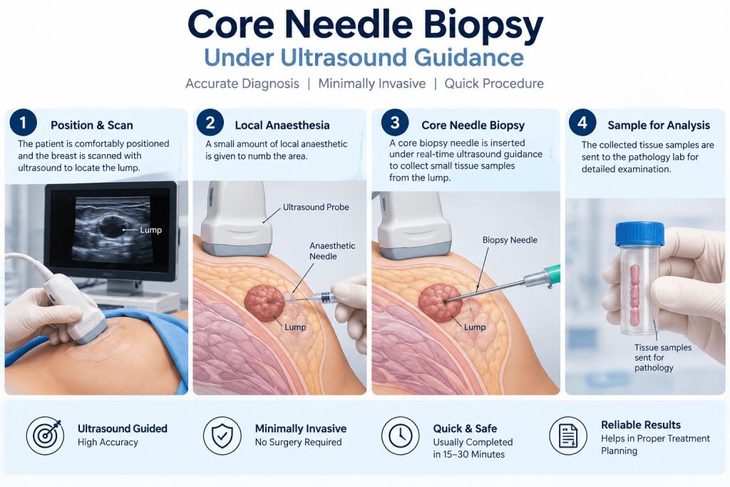
How It Works
- Imaging guidance — Using real-time ultrasound (or, for certain lesions, stereotactic mammographic or MRI guidance), the interventional radiologist precisely visualizes the lump throughout the procedure.
- Local anesthesia — A small amount of local anesthetic numbs the skin and breast tissue at the entry site.
- Sampling — A specialized, spring-loaded core needle is guided directly into the lump under continuous imaging visualization, removing several small cylindrical tissue samples (rather than fluid or loose cells, as with a fine-needle aspiration) — enough tissue for a pathologist to examine the actual architecture of the lesion under a microscope, not just individual cells.
- Marker placement — A tiny, imaging-visible marker clip is often placed at the biopsy site, so the exact location can be found again on future imaging if needed.
- Recovery — A small dressing is applied; the entire procedure typically takes well under an hour, and patients go home the same day with no stitches required.
- Also Read: Is It Normal to Have a Headache Every Day? Causes & Treatment | CVIC Indore
Preparing for the Procedure and What to Expect Afterward
Preparation is minimal compared with any surgical procedure: patients are generally asked to avoid blood-thinning medication in the days beforehand only if specifically advised, and to wear comfortable, easy-to-remove clothing on the day. No fasting is typically required, since only local anesthesia is used. After the procedure, mild tenderness, bruising, or minor swelling at the site is common for a few days and usually managed well with a cold compress and simple over-the-counter pain relief; strenuous upper-body exercise is generally best avoided for a day or two. Most patients are back to their normal daily routine, including work, the same day or the next.
Why This Matters: Accuracy Without Surgery
Published comparative data shows that core needle biopsy is almost as accurate as open surgical biopsy, while carrying a meaningfully lower risk of complications and leading to far fewer follow-up surgical procedures than the old surgery-first approach — because most patients now get a definitive answer without ever entering an operating theater. The one honest limitation worth knowing: because a core needle samples only part of a lesion rather than the whole thing (unlike surgical excision, which typically removes the entire mass), there is a small residual chance that an unsampled area of a large or complex lesion behaves differently than the sampled tissue — which is exactly why radiologists correlate biopsy results against the imaging appearance (concordance) and may recommend either surgical excision or continued monitoring in specific situations where the two don’t add up.
Core Needle Biopsy vs. Surgical (Excisional) Biopsy
| Factor | Core Needle Biopsy | Surgical Biopsy |
|---|---|---|
| Setting | Outpatient, imaging suite | Operating room |
| Anesthesia | Local only | Local, sedation, or general |
| Incision | None (needle puncture only) | Yes, with stitches |
| Scarring | Minimal to none | Visible surgical scar |
| Recovery | Same day, minimal restriction | Days to weeks |
| Tissue sampled | Representative core samples | Entire lesion (in most cases) |
| Diagnostic accuracy | Very high; almost equivalent to surgery | Considered definitive |
| Typical next step if benign | Monitoring or treatment as appropriate | Usually treatment-complete |
What Happens After Diagnosis
If the biopsy confirms fibroadenoma: Most patients have two reasonable paths — continued monitoring with periodic imaging (appropriate for smaller, stable, typical-appearing fibroadenomas), or active treatment if the fibroadenoma is large, growing, causing symptoms, or simply a source of ongoing anxiety the patient would rather resolve. For patients who want the lump treated rather than watched, non-surgical options such as image-guided ablation are available — CVIC Indore’s dedicated resources on fibroadenoma treatment cover that next step in detail for patients who reach this point.
If the biopsy raises any concern or confirms malignancy: The same image-guided biopsy pathway that diagnosed the problem also typically provides the tissue needed for further pathology testing (such as hormone receptor status) that guides oncology treatment planning — meaning the diagnostic step is not wasted time, but a direct, efficient bridge into appropriate specialist care without unnecessary delay.
Why Choose CVIC Indore for Non-Surgical Breast Lump Diagnosis
CVIC Indore (Center Of Vascular And Interventional Care) brings the same image-guided precision used across its interventional radiology practice to breast lump evaluation:
- Real-time ultrasound-guided biopsy expertise, ensuring the needle samples exactly the right target with continuous visual confirmation throughout the procedure.
- A same-day, walk-in-walk-out approach to tissue diagnosis, avoiding the anesthesia, incision, and recovery time associated with surgical biopsy.
- A structured, honest diagnostic process — clinical correlation, imaging characterization, and tissue sampling when indicated, rather than reflexively recommending a procedure for every lump or, conversely, reflexively reassuring without adequate evaluation.
- A clear pathway to treatment for patients whose fibroadenoma is confirmed and who wish to proceed with non-surgical treatment, without having to start the process over at a different facility.
Myths vs. Facts
Myth: If a lump doesn’t hurt, it’s probably nothing to worry about. Fact: Pain is an unreliable indicator either way — most early breast cancers are painless, and many benign lumps and cysts can be tender. Absence of pain should never delay evaluation of a new lump.
Myth: If a lump feels smooth and moves easily, it must be a fibroadenoma. Fact: While this is the classic description of a fibroadenoma, some cancers — particularly in younger women — can feel deceptively similar on examination. Touch alone cannot make this distinction reliably.
Myth: Getting a biopsy means the doctor already suspects cancer. Fact: A biopsy is often recommended specifically to confirm a benign diagnosis with certainty, or because a finding falls into an intermediate imaging category (BI-RADS 4) where the risk range is wide — not because malignancy is expected.
Myth: You need surgery to get a definitive breast lump diagnosis. Fact: Image-guided core needle biopsy provides diagnostic accuracy nearly equivalent to surgical biopsy for the large majority of patients, without an operating room, anesthesia beyond local numbing, or a surgical scar.
Myth: A “probably benign” (BI-RADS 3) result means no further action is needed. Fact: BI-RADS 3 findings are managed with structured short-interval follow-up imaging specifically because a small risk remains — skipping that follow-up defeats the purpose of the classification.
Frequently Asked Questions
1. I have a lump that feels exactly like what people describe as a fibroadenoma — do I still need imaging? Yes. Clinical impression alone is not considered adequate diagnosis under the triple assessment standard, since imaging reveals internal characteristics that touch cannot detect, and imaging findings directly determine whether biopsy is needed.
2. Is core needle biopsy painful? The area is numbed with local anesthetic beforehand, so most patients feel pressure rather than sharp pain during sampling. Mild soreness at the site for a day or two afterward is common and manageable with simple pain relief.
3. How long does it take to get biopsy results? This varies by pathology laboratory turnaround, but results are typically available within a few days to about a week.
4. If imaging says BI-RADS 3, why does my doctor want to just watch it instead of doing a biopsy right away? BI-RADS 3 reflects a low (under 2%) probability of malignancy based on classic benign imaging features. Structured short-interval follow-up imaging is a well-validated, evidence-based approach for this category, reserving biopsy for lesions that show any change.
5. Can a fibroadenoma turn into cancer? Standard fibroadenomas do not transform into cancer and do not meaningfully raise a woman’s future breast cancer risk. A rare, different type of tumor called a phyllodes tumor can sometimes mimic a fibroadenoma on initial impression, which is part of why tissue diagnosis matters for larger or changing masses rather than assuming every fibroadenoma-like lump is identical.
6. Will I have a scar after core needle biopsy? No incision is made — only a needle puncture — so visible scarring is minimal to none, unlike surgical biopsy, which requires an incision and stitches.
Final Thoughts
A new breast lump deserves a clear answer, not just reassurance or anxiety. The combination of clinical examination, imaging characterization using standardized BI-RADS reporting, and — when indicated — non-surgical tissue diagnosis through image-guided core needle biopsy gives patients and doctors a structured, evidence-based way to tell breast lump or fibroadenoma apart from something that needs urgent attention, without defaulting to either false reassurance or unnecessary surgery.
If you’ve found a new breast lump, the right next step is a proper clinical evaluation and imaging — not a search for symptoms online, and not indefinite worry. The team at CVIC Indore can guide you through that process and, if a biopsy is indicated, provide it without surgery.
Visit CVIC Indore
CVIC — Center Of Vascular And Interventional Care 403, Panama Tower, Gita Bhawan Road, near Crown Palace, South Tukoganj, Indore, Madhya Pradesh 452008
Phone: 0731 4675670 / +91-8103727728
Team: Dr. Alok K. Udiya (MD Radiology, Fellowship in Neuro Intervention Radiology, Switzerland), Dr. Shailesh Gupta (MD Radiology, PDCC in Interventional Radiology), Dr. Nishant Bhargava (MD Radiology, Fellowship in Neuro Intervention Radiology, Switzerland)
Disclaimer
This article is intended for general educational purposes only and does not constitute medical advice. Diagnosis of any breast lump requires in-person clinical examination and, where indicated, imaging and tissue sampling by a qualified physician or radiologist. If you have found a new breast lump or noticed a change in an existing one, please consult a doctor promptly for personalized evaluation.