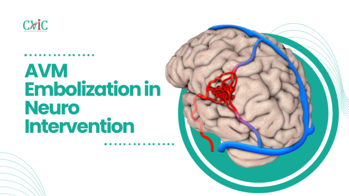
Arteriovenous Malformation (AVM) causes arteries and veins in the brain or spine to tangle, leading to abnormal blood flow. Neuro interventionists use AVM embolization to treat AVMs. This blog aims to provide a detailed overview of AVM embolization, including its purpose, procedure, risks, and recovery.
What is AVM Embolization?
Neuro interventional radiologists use AVM embolization as a minimally invasive procedure to reduce or eliminate blood flow to an AVM. During the procedure, a neuro interventional radiologist inserts a catheter into an artery, typically in the groin, and guides it to the location of the AVM in the brain or spine. Once the catheter is in place, the radiologist injects a glue-like substance or tiny particles called embolic agents into the AVM. These agents block the abnormal blood vessels, reducing the risk of bleeding and potentially causing the AVM to shrink over time.
Purpose of AVM Embolization in Neuro Intervention
The primary goal of AVM embolization is to reduce the risk of bleeding or rupture of the AVM, which can lead to serious complications such as stroke, brain damage, or even death. By blocking the abnormal blood vessels, AVM embolization helps to normalize blood flow in the affected area and reduce the risk of these complications.
Procedure in AVM Embolization
Preparation: Before the procedure, the patient will undergo imaging tests, such as angiography, to map the AVM and identify the best approach for treatment.
Anesthesia: The procedure is performed under local anesthesia, meaning the patient will be awake but will not feel any pain. In some cases, sedation may be used to help the patient relax.
Catheterization: A small incision is made in the groin, and a catheter is inserted into an artery and guided to the location of the AVM using real-time imaging guidance.
Embolization: Once the catheter is in place, the neuro interventional radiologist injects the embolic agent into the AVM, blocking the abnormal blood vessels and reducing blood flow to the area.
Monitoring: The radiologist will monitor the progress of the embolization using imaging techniques to ensure that the AVM is being effectively treated throughout the procedure.
Closure: After the procedure is complete, the catheter is removed, and the incision site is closed with stitches or a special closure device.
Risks and Complications
While AVM embolization is generally considered safe, like any medical procedure, it carries some risks, including:
Bleeding: There is a risk of bleeding at the catheter insertion site or within the brain or spine.
Stroke: In rare cases, the embolic agent can block blood flow to healthy brain tissue, leading to a stroke.
Infection: There is a small risk of infection at the catheter insertion site.
Allergic reaction: Some patients may have an allergic reaction to the embolic agent.
Recovery and Follow-up
After the procedure, hospital typically monitors patients for a few hours or overnight. Most patients can typically resume normal activities within a few days to a week, although they should typically avoid strenuous activities for a few weeks. Typically, healthcare providers may perform follow-up imaging tests to assess the effectiveness of the embolization and monitor the AVM over time.
Conclusion
AVM embolization is a valuable treatment option for patients with AVMs in the brain or spine. By blocking abnormal blood vessels, this minimally invasive procedure can reduce the risk of bleeding and other serious complications associated with AVM Embolization in Neuro Intervention. While it carries some risks, the benefits of AVM embolization can be life-saving for many patients.
Our Doctors
Dedicated IR Center for Vascular Problems in Madhya Pradesh
DR. SHAILESH GUPTA
MD, PDCC (INTERVENTIONAL RADIOLOGY) Consultant & Co-Director CVIC (Center Of Vascular & Interventional Care)
DR. ALOK KUMAR UDIYA
MD Radiology, PDCC (Neuro intervention Radiology), PDCC ( HPB Intervention Radiology) FINR (Switzerland) & EBIR
Endovascular Surgeon & Consultant Interventional Neuroradiologist at Care CHL Hospital, Indore Co-director CVIC( center for vascular and interventional care) https://interventionradiologyindore.com/
DR. NISHANT BHARGAVA
Consultant Intervention Radiologist
MD Radiology, PDCC ( Neuro intervention Radiology), FINR ( Fellowship in Neuro intervention Radiology)
Co-director CVIC(Center for Vascular and Interventional Care)
Contact Details
Phone no.
0731 4675670
+91 9827760073
Facebook
https://www.facebook.com/profile.php?id=100092538633553&mibextid=ZbWKwL
Instagram
https://instagram.com/cvic_center?igshid=ZGUzMzM3NWJiOQ==
Google My business
https://g.co/kgs/DrdV3T
YouTube
https://www.youtube.com/channel/UCP5TH5e4iQZkpDUgnLsgZhw
Pinterest
https://pin.it/5DzpX5Z
Twitter
https://x.com/cviccenter?t=01TclSrLFdu0K2re0Gs96w&s=08
LINKEDIN
https://www.linkedin.com/company/center-of-vascular-interventional-care/
Location –
Read More –
Carotid Stenting in Neuro Intervention – https://cvicvascular.com/carotid-stenting-in-neuro-intervention/
Aneurysm coiling in Neuro Intervention – https://cvicvascular.com/aneurysm-coiling-in-neuro-intervention/
Pre-operative Embolisation of Tumor in Neuro Intervention – https://cvicvascular.com/pre-operative-embolisation-of-tumor/


