Introduction
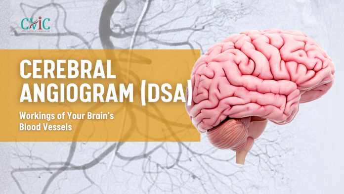
Our brains are one of the most complex and vital organs in the human body. They require a constant supply of oxygen and nutrients to function correctly. This supply is facilitated by an intricate network of blood vessels, known as the cerebral vascular system. Sometimes, issues with these blood vessels can arise, leading to potentially life-threatening conditions such as aneurysms, vascular malformations, or strokes. To diagnose and treat these conditions, doctors often turn to a specialized imaging technique called a Cerebral Angiogram, also known as Digital Subtraction Angiography (DSA). In this comprehensive guide, we’ll delve into the world of cerebral angiograms, explaining what they are.
Section 1: Understanding the Cerebral Vascular System
1.1 The Importance of Brain Blood Vessels
The brain is the control center of the body, managing all bodily functions and cognitive processes. It relies on a continuous supply of oxygen and nutrients delivered via blood vessels. The intricate network of arteries, veins, and capillaries that make up the cerebral vascular system is responsible for ensuring the brain receives these essential resources.
1.2 Common Cerebrovascular Conditions
Several conditions can affect the cerebral vascular system, leading to health problems. These conditions include:
- Aneurysms: Weak spots in blood vessel walls that can rupture, causing potentially fatal bleeding in the brain.
- Arteriovenous Malformations (AVMs): Abnormal tangles of blood vessels that can disrupt blood flow and cause hemorrhages.
- Stenosis: Narrowing of blood vessels, which reduces blood flow to the brain, potentially leading to stroke.
- Tumors: Abnormal growths that can press on or disrupt blood vessels in the brain.
Section 2: What is a Cerebral Angiogram?
2.1 Cerebral Angiogram Definition
A cerebral angiogram, or Digital Subtraction Angiography (DSA), is a diagnostic imaging procedure used to visualize the blood vessels in the brain.
2.2 The Purpose of Cerebral Angiography
Cerebral angiography serves several critical purposes:
- Diagnosis: It helps identify and assess various cerebrovascular conditions, such as aneurysms, AVMs, stenosis, and tumors.
- Treatment Planning: By providing detailed images of the blood vessels, doctors can plan surgical or interventional procedures to address the detected issues.
- Follow-up
Section 3: The Cerebral Angiogram Procedure
3.1 Preparing for a Cerebral Angiogram
Before the procedure, patients need to take several steps to ensure a successful and safe examination:
- Medical History: A thorough medical history is taken to assess the patient’s overall health and any existing conditions.
- Blood Tests: Blood tests may be necessary to evaluate blood clotting and kidney function.
- Medication Adjustments: Depending on the medications a patient is taking, adjustments may be needed prior to the procedure.
3.2 The Cerebral Angiogram Process
The actual procedure involves several key steps:
- Anesthesia: Cerebral angiograms are usually performed under local anesthesia, meaning the patient is awake but won’t feel pain in the area of the body being examined. Sedation may be provided to help the patient relax.
- Access Site: A small incision is made in the groin or arm, where a thin, flexible tube called a catheter is inserted into an artery.
- Contrast Injection: A contrast dye is injected through the catheter into the bloodstream, allowing the blood vessels in the brain to be visible on X-ray images.
- X-ray Imaging: X-ray images are taken in real-time as the contrast dye moves through the blood vessels. This process helps create a road map of the cerebral vascular system.
- Digital Subtraction: The DSA technique involves continuously subtracting a stationary image (without contrast) from a series of contrast-enhanced images. This results in clear and detailed images of the blood vessels.
- Image Review: The obtained images are reviewed by a radiologist to assess the health of the blood vessels and identify any abnormalities.
- Catheter Removal: Once the procedure is complete, the catheter is removed, and the access site is usually closed with sutures or a closure device.
3.3 Duration of the Procedure
The duration of a cerebral angiogram can vary but typically takes between 1 to 2 hours. This includes the preparation, the procedure itself, and some recovery time.
Section 4: Risks and Benefits of Cerebral Angiography
4.1 Benefits of Cerebral Angiography
Cerebral angiography offers several important benefits:
- Accurate Diagnosis: It provides high-resolution images that help accurately diagnose cerebrovascular conditions.
- Treatment Planning: It assists in planning surgical or interventional procedures.
- Real-time Imaging: The ability to capture real-time images is essential for assessing blood flow and identifying abnormalities.
- Minimal Invasive: It is a minimally invasive procedure, meaning it requires only small incisions and generally has a shorter recovery time compared to open surgery.
4.2 Risks and Complications
While cerebral angiography is generally safe, it carries certain risks and potential complications:
- Allergic Reactions: Some patients may be allergic to the contrast dye, which can lead to an allergic reaction.
- Blood Clots: There is a slight risk of developing a blood clot at the insertion site.
- Infection: Infections at the catheter insertion site are rare but possible.
- Stroke: In rare cases, the procedure can lead to a stroke, usually due to dislodging a blood clot or dislodged plaque from the artery wall.
Section 5: Recovery and Aftercare
5.1 Immediate Aftercare
After the procedure, patients are monitored in a recovery area for several hours to ensure there are no complications. During this time, medical staff will closely observe vital signs, including blood pressure and heart rate.
5.2 Post-Procedure Instructions
- Rest: Patients should rest and limit physical activity for a period following the procedure.
- Hydration: Staying well-hydrated helps flush the contrast dye from the body.
- Medications: Depending on the individual’s condition, medications may be prescribed.
- Follow-up: Patients typically have a follow-up appointment with their doctor to discuss the results and any necessary treatment.
Section 6: Conclusion and Future Outlook
In conclusion, cerebral angiography, or Digital Subtraction Angiography (DSA), is an invaluable diagnostic tool for assessing the health of the cerebral vascular system. It allows doctors to accurately diagnose cerebrovascular conditions, plan treatments, and monitor the success of interventions.
As medical technology continues to advance, we can expect further improvements in the safety and precision of cerebral angiography. New imaging techniques and minimally invasive procedures will further enhance our ability to protect and maintain brain health. By understanding the importance of procedures like cerebral angiography, we can take proactive steps to ensure the well-being of our most vital organ, the brain.
Our Doctors –
Dedicated IR Center for Vascular Problems in Madhya Pradesh
DR. SHAILESH GUPTA
MD, PDCC (INTERVENTIONAL RADIOLOGY) Consultant & Co-Director CVIC (Center Of Vascular & Interventional Care)
DR. ALOK KUMAR UDIYA
MD Radiology, PDCC (Neurointervention Radiology), PDCC ( HPB Intervention Radiology) FINR (Switzerland) & EBIR
Endovascular Surgeon & Consultant Interventional Neuroradiologist at Care CHL Hospital, Indore Co-director CVIC( center for vascular and interventional care)
DR. NISHANT BHARGAVA
Consultant Intervention Radiologist
MD Radiology, PDCC ( Neurointervention Radiology), FINR ( Fellowship in Neurointervention Radiology)
Co-director CVIC(Center for Vascular and Interventional Care)
Contact Details –
Phone no.
0731 4675670
+91 9827760073
Facebook
https://www.facebook.com/profile.php?id=100092538633553&mibextid=ZbWKwL
Instagram
https://instagram.com/cvic_center?igshid=ZGUzMzM3NWJiOQ==
Google My business
https://g.co/kgs/DrdV3T
YouTube
https://www.youtube.com/channel/UCP5TH5e4iQZkpDUgnLsgZhw
Pinterest
https://pin.it/5DzpX5Z
Twitter
https://x.com/cviccenter?t=01TclSrLFdu0K2re0Gs96w&s=08
LINKEDIN
https://www.linkedin.com/company/center-of-vascular-interventional-care/
Location –
Read More –
Laser Treatment for Varicose Veins: A Comprehensive Guide – https://cvicvascular.com/laser-treatment-for-varicose-veins-a-comprehensive-guide/
Understanding Breast Fibroadenoma: Causes, Symptoms, Diagnosis, and Treatment – https://cvicvascular.com/understanding-breast-fibroadenoma-causes-symptoms-diagnosis-and-treatment/
A Comprehensive Guide to the Treatment of Hypothyroidism – https://cvicvascular.com/a-comprehensive-guide-to-the-treatment-of-hypothyroidism/


